
Figure 1. Hierarchy of protein components in Yb body assembly.
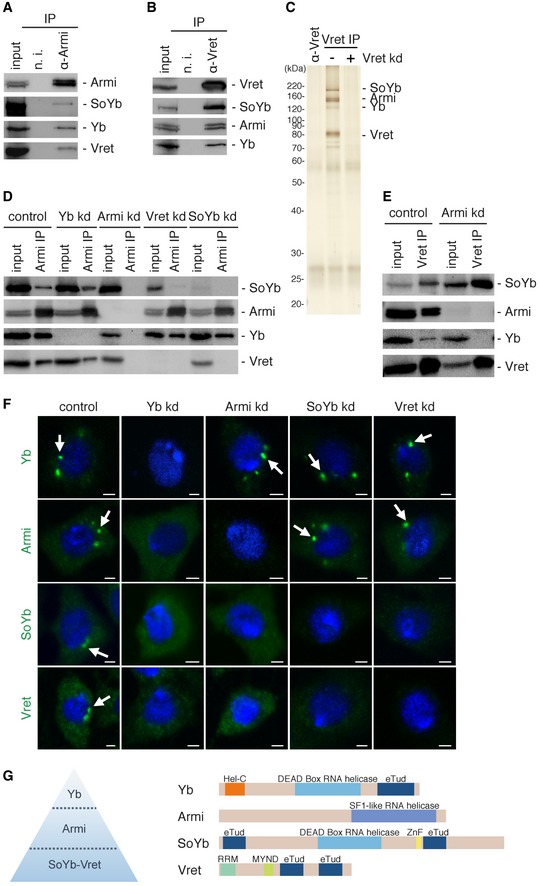
- The Armi complex immunoisolated from OSCs using anti‐Armi antibodies contains Yb, SoYb, and Vret.
- The Vret complex immunoisolated from OSCs using anti‐Vret antibodies contains Yb, Armi, and SoYb.
- Silver staining of protein components in the Vret complex in (B). The lane α‐Vret (left) contained only the anti‐Vret antibodies used in these experiments.
- Immunoprecipitation and Western blotting were performed in OSCs upon RNAi treatment (kd). Even without the association of Yb, Armi, SoYb, and Vret associated. Yb and Armi remained bound to each other upon SoYb or Vret depletion. The level of SoYb (input) decreased upon Vret depletion. Likewise, the level of Vret (input) decreased upon SoYb depletion.
- Upon Armi depletion, SoYb and Vret were still bound to each other but this heterodimer no longer associated with Yb.
- Immunofluorescence analyses in OSCs. Yb bodies disappeared upon Yb depletion, which led Armi, SoYb, and Vret to be localized in the cytosol. Yb bodies were detected in OSCs upon Armi depletion, but SoYb and Vret localized in the cytosol. Armi was detected at Yb bodies upon SoYb or Vret depletion. Yb bodies are indicated by white arrows. Nuclei are shown in blue. Scale bar: 2 μm.
- The hierarchy of Yb body assembly proposed in this study. Domain structures of Yb, Armi, SoYb, and Vret are shown on the right. Hel‐C, Helicase C‐terminal; eTud, extended Tudor; ZnF, zinc finger; RRM, RNA‐recognition motif; MYND, MYND‐type zinc finger.
Source data are available online for this figure.