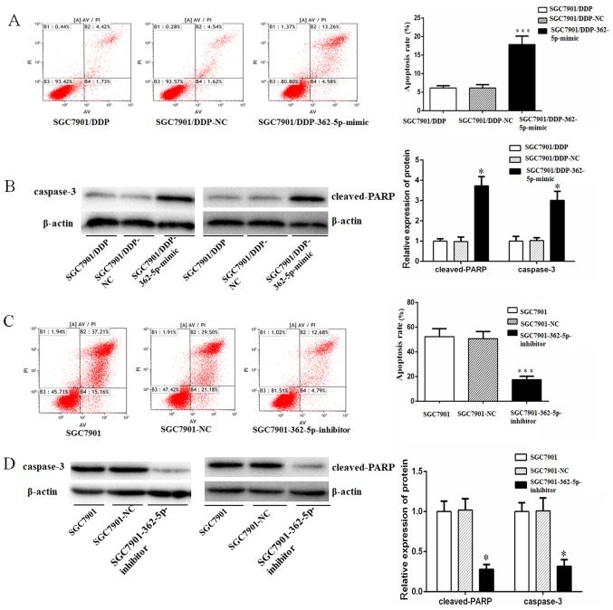
Figure 7.
miR-362-5p sensitizes gastric carcinoma cells to cisplatin-induced apoptosis. (A) Flow cytometry and (B) western blot analysis results demonstrated an increase in apoptotic rate (%) and the expression of apoptotic markers (caspase-3 and cleaved-PARP), respectively, following 48-h cisplatin treatment (final concentration of 0.8 µg/ml) in SGC7901/DDP cells transfected with miR-362-5p mimic compared with control cells. (C) Flow cytometry and (D) western blotting results indicated significant decreases in apoptotic rate and the expression of apoptotic markers (caspase-3 and cleaved-PARP), respectively, after 48-h cisplatin treatment (final concentration 0.3 µg/ml) in SGC7901 cells treated with miR-362-5p inhibitor compared with control cells. Columns indicate the mean of three independent experiments. *P<0.05; ***P<0.001. AV, annexin V-FITC; miR-362-5p, microRNA-362-5p; NC, negative control; PARP, poly-[ADP-ribose] polymerase; PI, propidium iodide.

