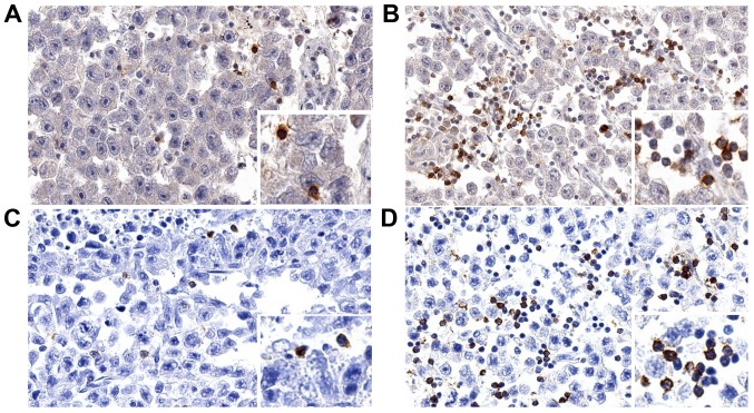
Figure 1.
(A and B) TIGIT and (C and D) PD-1 staining in seminomas. Examples of cases with low-level infiltration of (A) TIGIT+ and (C) PD-1+ immune cells and cases with high level of infiltration of (B) TIGIT+ and (D) PD-1+ immune cells. TIGIT, T cell immunoreceptor with Ig and ITIM domains; PD-1, Programmed Cell Death 1.

