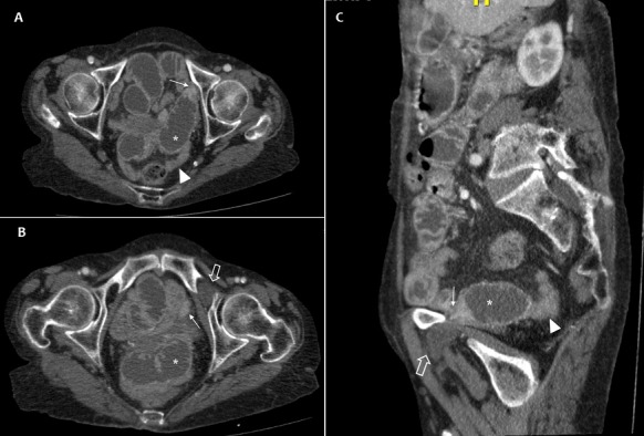
Figure 1.

(A) axial pelvic CT scan; (B,C) multiplanar reconstructed images through the left obturator canal, showed a dilated loop of small bowel (asterisk) upstream of a strangulated point (arrow) caused by a left obturator hernia (large arrow). Noted the collapsed loops down and colon (arrowhead)
