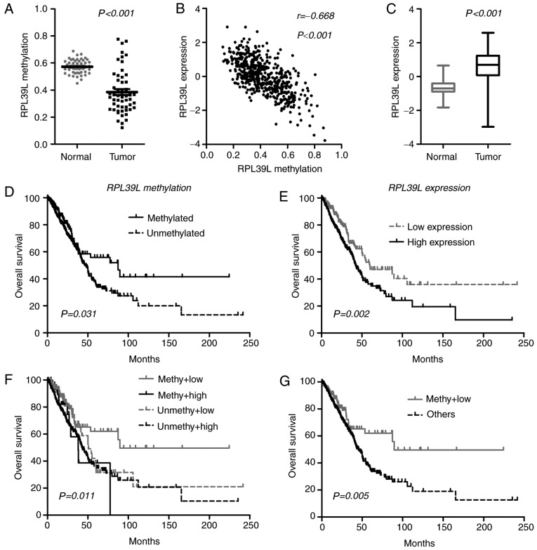
Figure 3.
Integration of DNA methylation and gene expression of RPL39L within The Cancer Genome Atlas samples. (A) Methylation status between matched lung adenocarcinomas and normal tissues. (B) Expression status between matched lung adenocarcinomas and normal tissues. (C) Pearson correlation between DNA methylation and RPL39L gene expression. (D) Patient classification on the basis of single-locus methylation levels of RPL39L. (E) Patient classification on the basis of the expression levels of RPL39L. (F) Patient classification on the basis of the combined assessment of DNA methylation and gene expression of RPL39L. (G) Patients with increased methylation and decreased expression of RPL39L experienced an improved survival time compared with that in the other subgroups. RPL39L, ribosomal protein 39 like; methy, methylated; unmethy, unmethylated.

