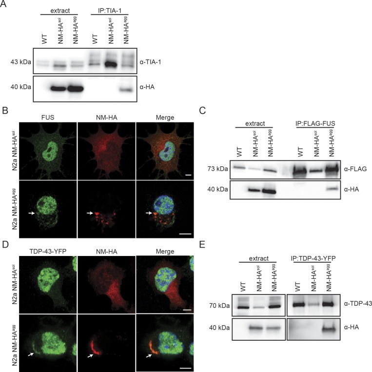
Figure S2. TDP-43 and FUS colocalize with NM-HA aggregates.
(A) IP of TIA-1 from lysates of wild-type N2a, N2a NM-HAsol, and N2a NM-HAagg cells using mAb anti-TIA-1, followed by SDS–PAGE and Western blot. Total cell lysate (extract) was loaded as control. TIA-1 was detected using pAb anti-TIA-1. NM-HA was detected using mAb anti-HA. (B) Immunofluorescence staining of N2a NM-HAsol and N2a NM-HAagg cells. NM was detected using mAb anti-HA (red), and endogenous FUS was detected using pAb anti-FUS (green). Nuclei were stained with Hoechst (blue). Scale bar: 5 μm. (C) Wild-type N2a, N2a NM-HAsol, and N2a NM-HAagg cells were transfected with a plasmid coding for FLAG-FUS. After 48 h, FUS was immunoprecipitated by using mAb anti-FLAG, followed by SDS–PAGE and Western blot. Total cell lysate (extract) was loaded as control. FUS was detected using mAb anti-FLAG, and NM-HA was detected using mAb anti-HA. (D) N2a NM-HAsol and N2a NM-HAagg cells were transfected with a plasmid coding for TDP-43-YFP and subjected to immunofluorescence staining after 48 h. NM was detected using mAb anti-HA (red), and TDP-43 was detected using pAb anti-TDP-43 (green). Nuclei were stained with Hoechst (blue). Scale bar: 5 μm. (E) Wild-type N2a, N2a NM-HAsol, and N2a NM-HAagg cells were transfected with a plasmid coding for TDP-43-YFP. After 48 h, TDP-43 was immunoprecipitated by using mAb anti-TDP-43, followed by SDS–PAGE and Western blot. Total cell lysate (extract) was loaded as control. TDP-43 was detected using mAb anti-TDP-43, and NM was detected using mAb anti-HA.
Source data are available for this figure.

