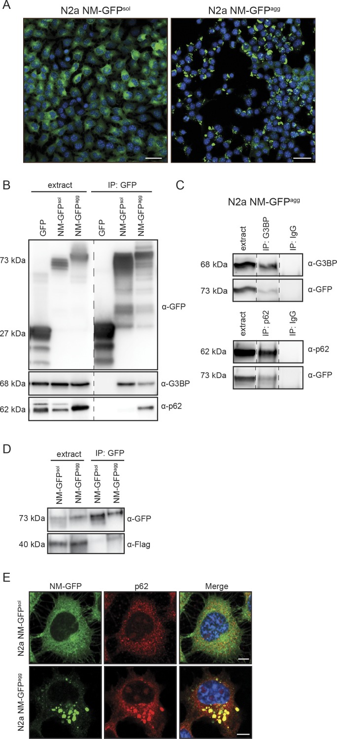
Figure S3. Interaction of NM-GFPagg with G3BP and p62.
(A) Bulk population of N2a cells stably expressing NM-GFP (N2a NM-GFPsol) and N2a clone stably propagating NM-GFP aggregates (N2a NM-GFPagg) (Hofmann et al, 2013). N2a NM-GFPagg cells were generated by exposing N2a NM-GFPsol cells to recombinant NM fibrils and subsequent limiting dilution cloning. Scale bar: 30 μm. (B) Validation of interaction of aggregated NM-GFP with G3BP and p62. NM-GFP in lysates of N2a NM-GFPsol or N2a NM-GFPagg cells was immunoprecipitated using GFP-trap magnetic beads. Co-immunprecipitated G3BP or p62 was detected by Western blot. Note that GFP was slightly degraded. Additional lanes were excised for presentation purposes (dashed line). (C) Control IP using unspecific IgG or anti-G3BP or anti-p62 IgGs and cell lysates of N2a NM-GFPagg. Additional lanes were excised for presentation purposes (dashed lines). (D) Pull-down of Flag-FUS transiently expressed in N2a NM-GFPsol and N2a NM-GFPagg cells. GFP-trap magnetic agarose beads were used for IP. (E) Colocalization of p62 with NM-GFP in N2a NM-GFPagg cells. N2a NM-GFPsol and N2a NM-GFPagg were stained with Hoechst and anti-p62. Scale bar: 5 μm.

