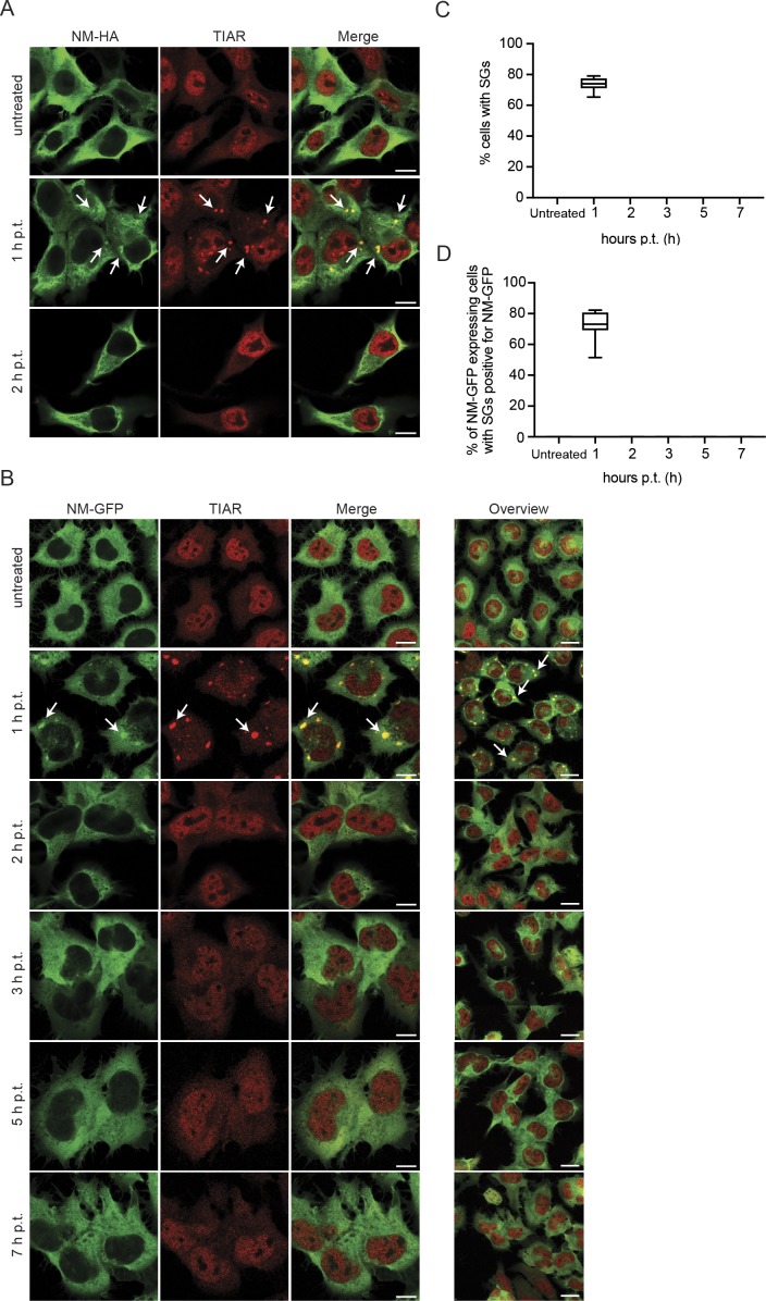
Figure S4. Association of NM-HA and NM-GFP with SGs is transient.
(A) N2a NM-HAsol cells were either left untreated or exposed to 0.5 mM arsenite for 30 min. Medium was replaced, and cells were incubated for up to 2 h posttreatment (p.t.). Fixed cells were stained using anti-HA and anti-TIAR antibodies. Scale bar: 10 μm. (B) N2a NM-GFPsol cells exposed to 0.5 mM arsenite for 30 min. Upon replacement of medium, cells were allowed to recover and fixed up to 7 h p.t. The panels on the left show representative individual cells. The panel on the right shows an overview of treated cells (scale bars: 10 and 20 μm, respectively). Arrows indicate SGs. (C) Percentage of cells of (B) with TIAR-positive SGs (only red channel). Treatment was performed in triplicates. For each time point, three images per independent experiment were analyzed. At least 400 cells per time point for each experiment were analyzed. (D) Percentage of NM-GFP expressing cells with SGs and colocalization of NM-GFP with SGs. Images of (B) were quantified. At least 300 cells per time point for each experiment were analyzed.

