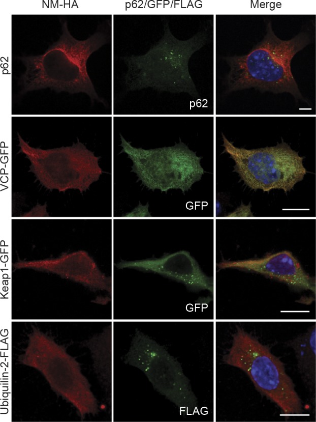
Figure S5. N2a NM-HAsol cells were left untreated (top row, for p62 staining) or transfected with constructs coding for VCP-GFP, Keap1-GFP, and Ubiquilin-2-FLAG and subjected to immunofluorescence staining 48 h posttransfection.

NM-HA was stained using mAb anti-HA (red), p62 was detected using mAb anti-p62 (green), and FLAG was stained using mAb anti-FLAG (green). GFP is shown in green. Nuclei were stained with Hoechst (blue). Scale bar: 5 μm.
