Fig. 1.

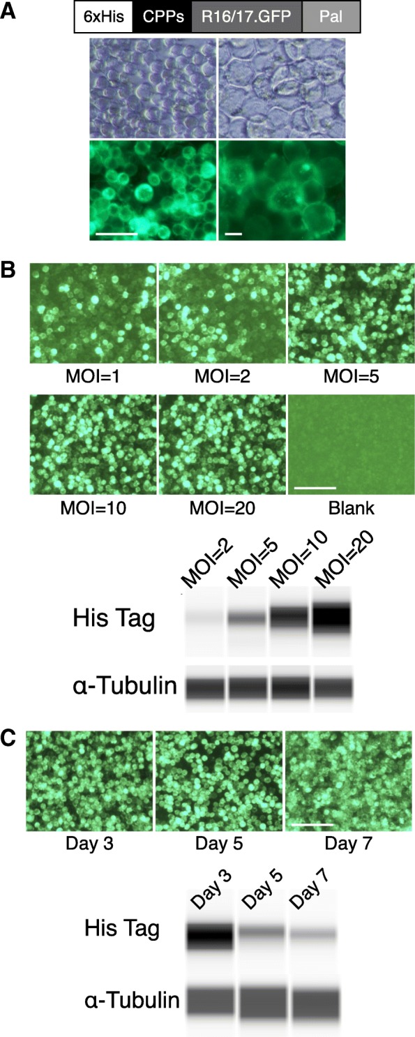
Dystrophin R16/17.GFP.Pal protein is expressed in insect cells and localized at the cell membrane. a Configuration of recombinant dystrophin R16/17.GFP.Pal protein and R16/17 protein expression in insect cells. Top panel: light microscopy; Bottom panel: fluorescence microscopy for GFP signal; Left panel: 10X; Right panel: 20X. The dystrophin R16/17.GFP.Pal protein was successfully expressed in the baculovirus-insect cell system and localized at the cell membrane. Left scale bar = 50 μm; Right scale bar = 10 μm; b Optimal MOI was determined for R16/17 protein production. The GFP signal was observed on day one after baculoviruses infection of High Five insect cells with different MOIs. The expression level from different MOIs was examined by western blot with an anti-His tag antibody, and α-tubulin serves as the loading control. Scale bar = 50 μm; c Optimal harvesting time was determined for R16/17 protein production. High Five cells were infected with baculoviruses (MOI = 20). The GFP signal and the expression of R16/17.GFP protein were examined 3, 5 and 7 days after virus infection by fluorescence microscopy and western blot with an anti-His tag antibody, respectively. The level of α-tubulin is used for the loading control. The highest protein expression was found 3 days after virus infection. Scale bar = 50 μm. Western blot was performed with ProteinSimple WES capillary western blot system
