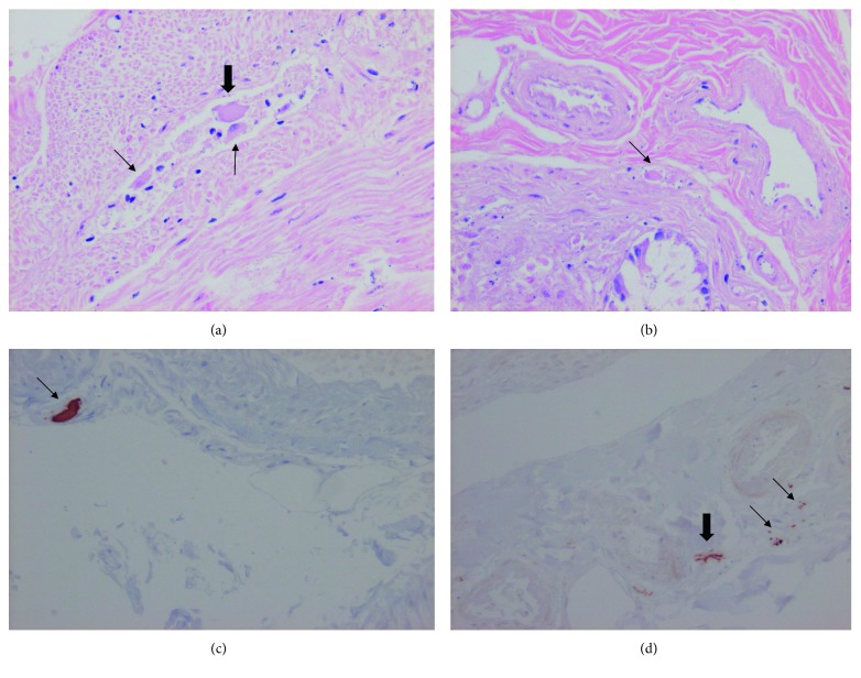
Figure 1.
(a) Myenteric shrunken plexus with one normal-sized neuron without nucleus (thick arrow) and two small atrophic/pyknotic neurons (thin arrows) (hematoxylin and eosin staining). (b) Submucosal plexus with one single atrophic neuron (arrow) (hematoxylin and eosin staining). (c) A large α-synuclein immunostaining of a submucosal plexus (arrow). (d) Smaller α-synuclein immunostaining of two submucosal plexa (thin arrows) and one axon (thick arrow) (magnification ×200).

