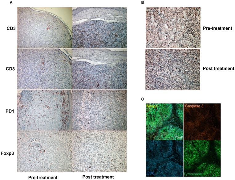
Figure 2.
Immunohistochemical staining for CD3, CD8, PD1, and Foxp3 before and after a single administration of nivolumab with ipilimumab (A). Immunohistochemical staining for PD-L1 before and after a single administration of nivolumab with ipilimumab (B). Immunofluorescence staining of CD8 (cytotoxic T cells: blue), caspase 3 (apoptotic cells: orange), and tyrosinase (melanoma cells: green) (C).

