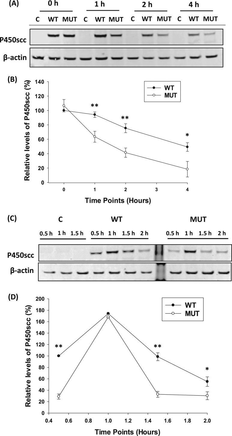
Figure 2.
Comparison of P450scc stability between WT and p.E314K variant. (A) Representative Western blot analysis of P450scc expression after WT or mutant (MUT) vector transfection for 48 hours in HEK293T cells followed by 25 µM CHX treatment of the indicated time points. A mock transfection without DNA was performed as a control. (B) Quantification of P450scc levels normalized to actin and expressed as relative to the WT 0 hour time point showed impaired protein stability. (C) Representative Western immunoblot of P450scc expression after transfection with WT or MUT vectors for 48 hours, followed by CHX treatment. After 4 hours, HEK293 cells were supplemented with fresh media, and lysates were collected at the indicated time points. (D) Quantification of P450scc expression normalized to actin and expressed as relative to the WT 0.5 hour time point after the CHX chase assay showed a decrease in protein levels. Data are shown as mean ± SD from three independent experiments. *P < 0.01; **P < 0.001. Abbreviation: C, control.

