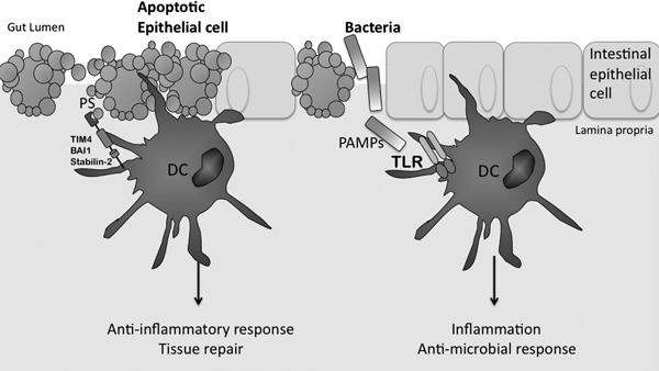
Figure 1.

Inflammatory infection and immunosuppressive apoptosis. At steady‐state, DCs phagocytose apoptotic cells during normal tissue turnover. This figure depicts the recognition of apoptotic epithelial cells by DCs at the epithelial surface. Here, the apoptotic cell expresses the eat me signal PS, which is recognized by PRRs expressed on the cell surface of DCs, such as TIM‐4, stabilin‐2, and BAI‐1. It has been shown that the regulatory cytokine TGF‐β is induced in response to uptake of apoptotic cells, and as such, this type of phagocytosis is known as noninflammatory. In contrast, microbial pathogens, as illustrated by bacteria here, activate TLRs on the surface of DCs by presenting ligands known as PAMPs, which are associated with particular bacteria. In this case, activated DCs produce proinflammatory cytokines, such as IL‐12, IL‐6, and TNF‐α, ultimately inducing an inflammatory, antimicrobial response required to fight the infection.
