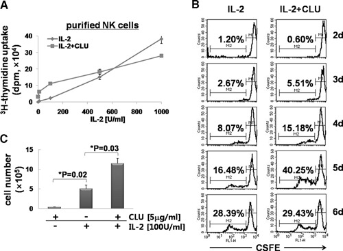
Figure 2.

CLU synergizes with IL‐2 for the proliferation of NK cells. (A) Purified NK cells were plated at 1 × 10⁵ cells/well and incubated for 3 days with various concentrations of IL‐2 (10, 100, 500, and 1000 U/ml) in the presence or absence of 5 μg/ml CLU. 3H‐Thymidine (1 μCi) was added for an additional 12 h to count radioactivity incorporated into the DNA. (B) Purified NK cells (5×10⁵) were labeled with 5 μM CFSE for 10 min, and cell division was analyzed by FACS, as described in Materials and Methods. Numbers in each graph indicate the percentage of cells undergoing cell division. FL1‐H, Fluorescence 1‐height. (C) Purified NK cells (1×10⁶) were cultured in the presence or absence of 5 μg/ml CLU and/or 100 U/ml IL‐2 for 6 days. Live cells were counted using a hemocytometer. The data shown are representatives of a minimum of 3 independent experiments. Error bars represent sd (*P<0.05).
