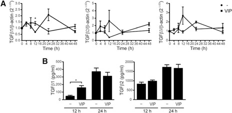
Figure 3.

VIP induces the expression of TGF‐β in trophoblast cells. (A) A kinetic analysis of TGF‐β1, ‐β2, and ‐β3 expression on Swan‐71 cells in the absence or presence of VIP (100 nM) by qRT‐PCR. Results are expressed as the fold change relative to β‐actin (2− ΔΔ Ct ± sem) and corresponded to 4 independent experiments. *P < 0.05, Student's t test. (B) In parallel, supernatants were collected, and Luminex quantification was performed for the 3 TGF‐β isoforms after 12 and 24 h of VIP treatment. Results are expressed as the mean pg/ml ± sem of TGF‐β secretion and are representative of 3 independent assays. *P < 0.05, Student's t test. TGF‐β3 was not detected under these conditions.
