
Figure 1.
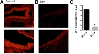
Sphingosine expression in bronchial epithelial cells of burn‐ and sham‐injured mice. Representative fluorescent staining of bronchial epithelial cells for Cy3‐coupled anti‐SPH antibodies from (A) sham and (B) burn‐injured mice are depicted. The images show the range of response of SPH levels before (control) after burn injury (burn). (C) Quantification of SPH expression (n = 4 mice/group). *P < 0.05 vs. sham group.