
Figure 4.
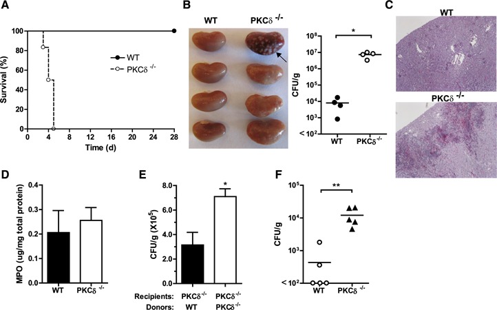
PKC‐δ is required for resistance to fungal infection. (A) PKC‐δ−/− mice and their WT cohorts (n = 8 in each) were infected intravenously with 5 × 10⁴ C. albicans and monitored daily for survival. P = 0.0002 (log‐rank test). (B) Kidneys were harvested at d 3 after infection for gross morphology and visual inspection of abscesses (arrow) (left panel) and for quantitation of fungal burden in kidney homogenates (right panel). Each symbol represents data from an individual animal. (C) A representative photomicrograph of periodic acid–Schiff (PAS)‐stained kidney sections from wild‐type and PKC‐δ−/− mice is shown. Within kidney tissues from PKC‐δ−/− mice, large collections of neutrophils (abscess formation) surrounded by mixed inflammatory cells are present with abundant fungal forms, which are virtually absent in kidneys from wild‐type mice. (D) Kidneys harvested from mice at 16 h after infection were homogenized, and MPO activity was determined and normalized to total protein content. Means ± sem of results are given. (E) PKC‐δ−/− mice were injected with WT or PKC‐δ−/− neutrophils followed by C. albicans inoculation; 20 h later, the colony‐forming unit in kidney homogenates was determined. (F) Mice were infected intranasally with 107 A. fumigatus conidia, and 2 d later the lung was harvested. The colony‐forming unit per gram of tissue was quantitated, and data are reported as in (B). *P < 0.05; **P < 0.01.