
Figure 4.
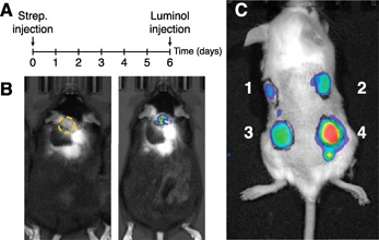
In vivo monitoring of MPO activity in mice. (A) Experimental design of the imaging study is shown. (B) A representative image of a mouse 6 d after s.c. injection of Streptococcus pyogenes (3 × 10¹⁰ CFU in phosphate‐buffered saline) in the upper back of the animal shown in white light (right image) and bioluminescence imaging (left image). A yellow dotted circle indicates the region of interest. (C) An example of the hydrogen peroxide (H2O2)‐dependence of MPO in vivo activity is shown. Albino C57BL/6 mice injected with 180 nM of MPO with either 400 μM of H2O2 (site 1), 2 mM of H2O2 (site 2), 4 mM of H2O2 (site 3), and 40 mM of H2O2 (site 4) were added to the matrigel (100 μl) before s.c. implant. (B–C) To visualize the MPO activity, an i.p. injection of luminol (Sigma‐Aldrich, 3 mg/kg) was given 10 min before luminescence imaging using the IVIS Lumina XRMS system (PerkinElmer, Waltham, MA, USA). The Institutional Animal Care and Use Committee of Auburn University approved all animal procedures.