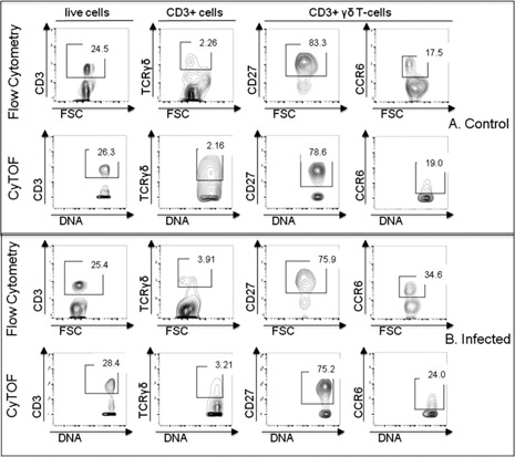
Figure 2.

Comparison of conventional flow cytometry to CyTOF. Male mice were infected intranasally with 1 × 10⁶ CFUs of S. pneumoniae (n = 4). Healthy, uninfected mice served as controls (n = 4). Spleen cells were harvested 1 d after infection and stained for either flow cytometry or CyTOF using anti‐CD3, anti‐TCRγδ, anti‐CD27, and anti‐CCR6 antibodies. Numbers in gates represent percentage of parent population as indicated in the row titles. (A) Biaxial plots of cells from control mice. (B) Plots of cells from mice at 1 d after infection. Representative plots demonstrate that conventional flow cytometry and CyTOF staining showed comparable staining profiles. Data are representative of 2 independent experiments. FSC, forward scatter.
