
Figure 2.
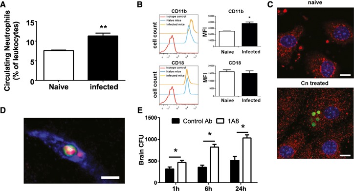
Enhanced expression of Mac‐1 and ICAM‐1 stimulated by Cn. (A) Numbers of neutrophils in the peripheral blood of mice (n = 4) were enhanced, 3 h after i.v. infection with 20 × 106 Cn (gated on CD45+ cells). (B) CD11b (upper), but not CD18 (lower), expressed on circulating neutrophils was elevated significantly in mice (n = 5), 3 h after i.v. infection with 20 × 106 Cn (gated on CD45+Ly6G+ cells). (C) ICAM‐1 staining was observed, to a greater extent, on mouse endothelial cell line (bEnd.3) treated with Cn in vitro (right) compared with untreated control (naïve; left). ICAM‐1, red; Cn, green; nuclei, blue. Original scale bars, 10 µm. (D) ICAM‐1 (red) intensity was increased around the arrested site of Cn (green) in the brain microvasculature (blue), 3 h after i.v. infection with 20 × 106 Cn. Original scale bar, 5 µm. (E) Brain CFU of mice (n = 3–4)‐treated anti‐Ly6G antibody (to deplete neutrophils) or control antibody at various time points after i.v. infection with 5 × 10⁴ Cn. *P < 0.05; **P < 0.01. Data are presented as means ± sem. Data are representative of 2–3 independent experiments.