
Figure 1.
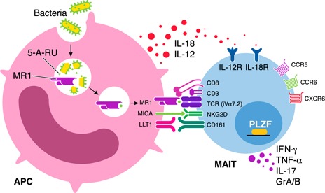
Mechanisms involved in the activation of MAIT cells. MAIT cells express a semi‐invariant TCR‐α chain, including an iVα7.2 segment combined with restricted Jα segments (Jα33, Jα12, or Jα20) and limited Vβ repertoires in humans. In addition, human MAIT cells express high levels of CD161, IL‐18Rα, the transcription factor PLZF, and the chemokine receptors CCR5, CCR6, and CXCR6. Mature MAIT cells are defined as CD161hiPLZFhiIL‐18Rα+iVα7.2+γδ−CD3+ lymphocytes. MAIT cells recognize unstable pyrimidine intermediates, formed by the nonenzymatic condensation of 5‐A‐RU, an early intermediate of vitamin B2 (riboflavin) synthesis, with glyoxal or MeG, derived from other metabolic pathways, presented by the highly evolutionarily conserved MR1 on APCs. This interaction leads to activation of the MAIT cell. After activation, MAIT cells can promptly kill infected cells, inhibit intracellular microbial growth, and produce proinflammatory cytokines, including IFN‐γ, TNF‐α, and IL‐17. It is noteworthy that MAIT cells can also be activated via exposure to the cytokines IL‐12 and IL‐18 in a TCR‐independent manner. MAIT cells also express high levels of NKG2D, which has a role as a cytotoxicity coreceptor.