
Figure 1.
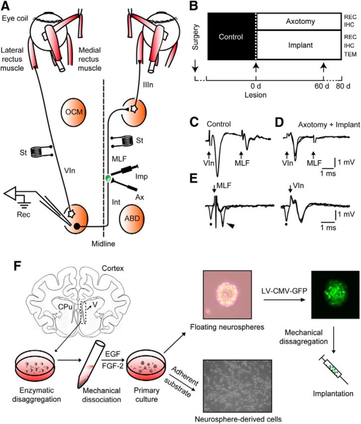
Experimental design and cellular identification. A, Diagram of connections of abducens (ABD) motoneurons and internuclear neurons (Int). Bipolar stimulating (St) electrodes were implanted in the VIn and in the MLF, respectively. The right MLF was sectioned (Ax) and implanted with neural progenitor cells (Imp). Recordings (Rec) were performed in the left ABD nucleus. B, Diagram of the time course of treatments and experimental procedures. IHC, Immunohistochemistry; REC, recordings; TEM, transmission electron microscopy. C, Antidromic field potentials obtained in ABD before lesioning by electrical stimulation of the VIn and MLF, respectively. D, Same as B, but after lesioning the MLF. E, Collision testing of an ABD Int. Left traces, Antidromic activation (arrowhead) of the Int from the MLF failed when conditioned by a prior spontaneous spike (dot). Right traces, Absence of collision of the orthodromic spike (dot) when the electrical shock was applied to the VIn. F, The subventricular zones were dissected aseptically from cat [postnatal day 3(P3) to P5] coronal sections at the lateral ventricle level (V), and enzymatically and mechanically disaggregated. The resulting cell suspension was cultured, and cells grew as floating neurospheres. On a poly-d-lysine adherent substrate, neurosphere-derived cells adhered to the dish and differentiated. Alternatively, neurospheres were transduced with lentiviral vectors containing a reporter gene for green fluorescent protein expressed under the control of a cytomegalovirus (CMV) promoter [lentiviral (LV)-CMV-GFP] and implanted in axotomized cats.