Figure 2.
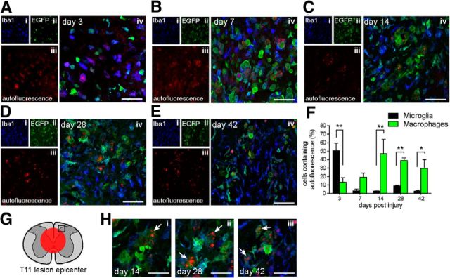
Phagocytosed material persists in peripherally derived macrophages but appears only transiently in microglia after SCI. A–E, Representative confocal images of microglia (Iba1+/EGFP−) and peripherally derived macrophages (Iba1+/EGFP+) containing phagocytosed, autofluorescent material (λ = 594 nm) at 3, 7, 14, 28, and 42 d after SCI as indicated. Single-plane confocal fluorescent images of Iba1 (i), EGFP (ii), and autofluorescence (iii) are shown on the left, and merged right (iv). Scale bars, 50 μm. F, Number of microglia containing autofluorescent material are greatly reduced after the initial peak at 3 d, in contrast to EGFP+ peripherally derived macrophages, which continue to contain phagocytosed debris 42 d after SCI. G, Schematic of cross section of spinal cord showing approximate site of contusion injury (red circle) and region used for quantification (black rectangle). H, Higher magnification confocal images of weakly EGFP+ cells highly laden with autofluorescent material (arrows), possibly indicating dead or dying cells at 14 (i), 28 (ii), and 42 d (iii) after SCI. Scale bar, 25 μm. Values expressed as mean ± SEM; *p < 0.05, **p < 0.01, n = 3–5.

