Figure 3.
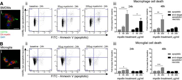
Peripheral macrophages are more susceptible than microglia to cell death after phagocytosis of myelin, in vitro. Fluorescent images showing BMDMs (Ai) and primary microglia (Bi) take up DiI-labeled myelin, in vitro. Aii, Bii, Representative FACS plots of CD45+ cells labeled for AnnexinV (apoptotic marker) and efluoro-780 (viability dye). Plots show level of apoptotic cell death (FITC-AnnexinV-positive, efluoro-780 viability dye positive; bottom right quadrant) and end-stage apoptosis/necrosis (efluoro-780 viability dye positive; top two quadrants) with increasing amounts of myelin added for 24 h. Aiii, Peripheral-derived macrophages show significant increase in apoptosis after treatment with 200 μg/ml myelin (24 h, 8.7-fold and 48 h, 6.1-fold). Biii, Primary microglia show much lower levels of apoptotic cell death in cultures treated with 200 μg/ml myelin (24 h, 1.7-fold and 48 h, 2.8-fold). End-stage apoptosis/necrosis increased 5.3-fold in peripherally derived macrophages at 24 h (200 μg/ml myelin), while necrotic cell death was not seen at either time point in primary microglia. Values expressed as mean ± SEM; *p < 0.05, **p < 0.01, ***p < 0.0001. n = 3–4. Scale bar, 20 μm.

