
Figure 7.
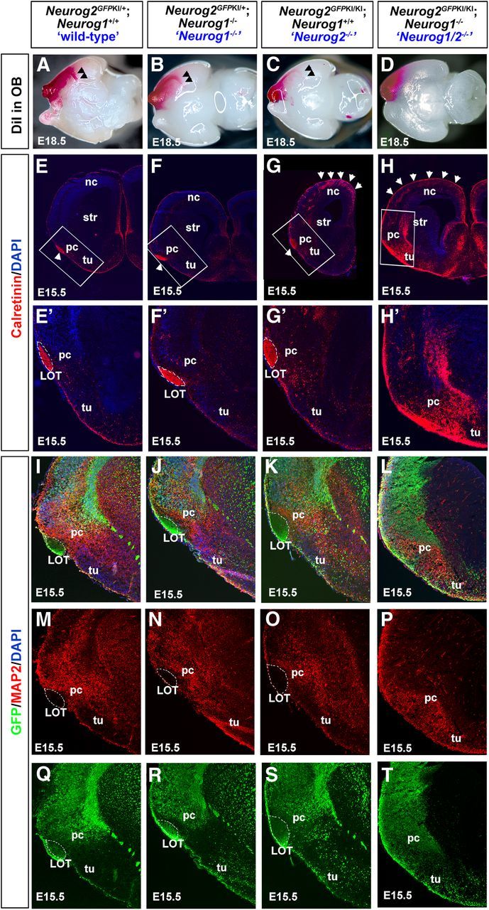
LOT formation is perturbed in the Neurog1/2−/− piriform cortex. A–D, LOT axons were traced by inserting a DiI crystal into E18.5 wild-type (A), Neurog1−/− (B), Neurog2−/− (C), and Neurog1/2−/− (D) OBs. Arrowheads mark the ventromedial turn of the forming LOT, which fails to form in Neurog1/2−/− embryos. E–H', Labeling of OB mitral cell axons in the LOT with anti-calretinin in E15.5 wild-type (E, E'), Neurog1−/− (F, F'), Neurog2−/− (G, G'), and Neurog1/2−/− (H, H') piriform cortices. Blue is a DAPI counterstain. E'–H' are higher-magnification images of boxed areas in E–H. Arrowheads in G and H mark supernumerary calretinin+ interneurons in Neurog2−/− and Neurog1/2−/− neocortices, respectively. I–T, Expression of GFP (green) and MAP2 (red) in E15.5 Neurog2GFPKI+/− heterozygotes (“wild-type”; I, M, Q), Neurog1−/− mutants carrying one copy of the Neurog2GFPKI allele (J, N, R), Neurog2GFPKI/KI mutants (K, O, S), and Neurog1−/−;Neurog2GFPKI/KI double mutants (Neurog1/2−/−; L, P, T). Blue is a DAPI counterstain. The dotted white line outlines the LOT. nc, Neocortex; pc, piriform cortex; str, striatum; tu, olfactory tubercle.