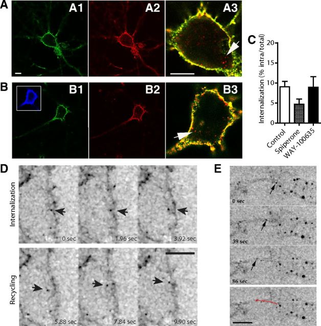
Figure 4.
Traffic of BBS-Flag-5-HT1AR in a transduced neuron. A, B, Immunostaining of hippocampal (A) and serotonergic raphe (B) neurons expressing BBS-Flag-5-HT1AR. BBS-Flag-5-HT1ARs localized at the plasma membrane were labeled in green (A1, B1), total BBS-Flag-5-HT1AR in red (A2, B2), and internalized BBS-Flag-5-HT1ARs were identified on merge picture (A3, B3) by red labeling (arrows) compared with yellow plasma membrane BBS-Flag-5-HT1AR. Serotonergic raphe neurons were labeled with anti 5-HT antibodies after permeabilization (B1, blue). C, Quantification of internalized BBS-Flag-5-HT1AR in serotonergic raphe neurons in control conditions and after exposure to spiperone or WAY-100635. D, E, Inverted monochrome images from time-lapse imaging of hippocampal neurons expressing BBS-Flag-5-HT1AR. D, Single events of internalization from plasma membrane and recycling in BBS-Flag-5-HT1AR-transduced neuron during a time lapse of 9.90 s (arrow). E, Intracellular anterograde traffic of BBS-Flag-5-HT1AR along proximal dendrites. Red line represents the traveled distance of BBS-Flag-5-HT1AR-positive vesicle (arrow) in 86 s. Scale bars: 5 μm.

