Figure 1.

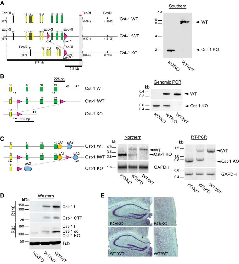
Characterization of a Cst-1 KO mouse. A, Left, Schematic representation of the Southern blot analysis of EcoRI-digested genomic DNA with a probe downstream of exon 19 of calsyntenin-1. Right, Southern blot demonstrating the presence of a sole 1.8 kb fragment in the homozygous Cst-1 KO mice, whereas WT mice contained the 8.7 kb fragment. B, Left, Schematic representation of the PCR analysis of genomic DNA as a complement to the Southern blot analysis. Right, PCR with primers a and b, the former located within the floxed segment, resulted in a 225 bp amplicon from the WT allele (right, top). Primers c and d were placed up and downstream, respectively, of the floxed segment and thus resulted in a 560 bp amplicon in the Cst-1 KO allele (right, bottom). C, Left, Schematic representation of the Northern blot analysis of polyadenylated RNA. Middle, The Northern blot demonstrates the presence of a stable truncated RNA transcript from the Cst-1 KO allele, indicating that the polyadenylation site is located downstream of the loxP site (pA2; left). Right, RT-PCR with primers e and f, located within exon 16 and at pA2, respectively, confirmed the presence of a Cst-1 KO transcript that is ∼0.7 kb shorter than the WT transcript. D, Immunoblots with antibody R140 raised against the cytoplasmic segment of calsyntenin-1 demonstrate the absence of the functionally active full-length transport form of calsyntenin-1 in homozygous Cst-1 KO mice together with an absence of its CTF. Antibody R85 against the N-terminal cadherin-like domains detected a truncated form of calsyntenin-1 in homozygous and heterozygous Cst-1 KO mice that is slightly shorter than the ectodomain in WT mice. E, Normal gross morphology of homozygous Cst-1 KO hippocampus (left) and cortex (right) revealed by Nissl staining. CTF, C-terminal fragment; ec, ectodomain; f, full-length; fWT, floxed wild-type; pA, polyadenylation site; Tub, tubulin.
