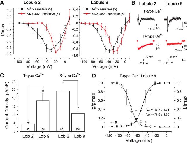
Figure 1.
Cerebellar granule cells express Cav3 calcium current. A, Comparisons between the mean I–V plots of calcium current in lobule 2 and 9 granule cells. Cav3 current was identified as the component sensitive to block by 300 μm Ni2+ (in the presence of 30 μm Cd2+) and R-type current as that blocked by 200 nm SNX-482 (in the absence of Cd2+). B, Representative T-type and R-type calcium currents in lobule 2 and lobule 9 granule cells. C, Bar plots of the mean density of T-type and R-type calcium currents isolated as in A and B for lobule 2 and 9 granule cells for a step from −110 to −30 mV. D, Superimposed mean conductance- and voltage-inactivation plots for T-type calcium current in lobule 9 granule cells. Sample numbers for mean values are shown in parentheses. *p < 0.05, Student's paired t test.

