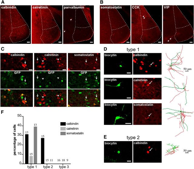
Figure 5.
Expression of interneuron markers in GABAergic neurons. A, B, Photomicrographs show MeA cell immunoreactivity for calbindin, calretinin, and parvalbumin and neuropeptides somatostatin, CCK, and VIP. Calbindin-ir (A, left), calretinin-ir (A, middle), and somatostatin-ir cells (B, left) were found in the MePV, whereas parvalbumin (A, right), CCK (B, middle), and VIP (B, right) neurons were largely absent. Arrows indicate immunoreactive cells for these markers in the neighboring regions: parvalbumin in the basomedial amygdala (A, right), CCK in the MePD (B, middle), and VIP in the cortical amygdala (B, right). C, Many calbindin-ir and almost all somatostatin-ir cells in the MePV were GFP+; calretinin was mostly found in GFP− cells. Photomicrographs represent MePV-immunoreactive cells for calbindin (top left), calretinin (top middle), and somatostatin (top right), expression of GFP (middle), and their colocalization (bottom). Arrows indicate GFP+ cells immunoreactive for these markers. Arrowheads indicate calretinin-ir cells that did not express GFP. D, All three markers were found in Type 1 neurons, whereas only calbindin was found in Type 2 cells (E). Reconstructed morphology of each cell is shown on the right panel (D, E). F, Percentage distribution of interneuron markers among electrophysiologically identified GFP+ neurons. Type 3 cells were not immunoreactive for any of the three markers. Numbers on top of bars indicate total number of cells that were processed in each group. M, Medial; V, ventral. Scale bars: A, B, 100 μm; C–E, 20 μm.

