
Figure 7.
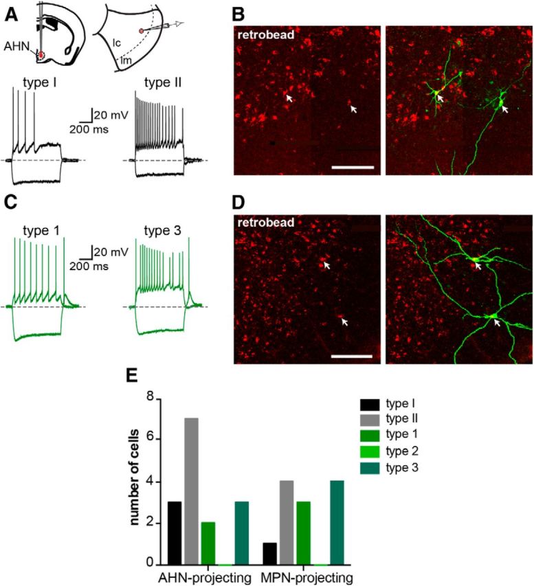
MePV neurons of different classes project to the same regions of the hypothalamus. A, Both types of GFP− cells project to AHN. Red retrobeads were injected into AHN, and recordings were made from GFP+ and GFP− retrogradely labeled neurons in the MePV (schematic). Traces represent hyperpolarization (bottom traces) and depolarization (top traces) responses of two retrogradely labeled AHN-projecting GFP− cells, identified as Type I (left) and Type II cells (right). B, Corresponding GFP− biocytin-filled neurons are shown. Photomicrographs represent retrogradely labeled neurons containing the red retrobead injected in AHN (left) and colocalization with biocytin-filled neurons (right). C, Both Type 1 and Type 3 GFP+ cells project to AHN. Traces represent hyperpolarization (bottom traces) and depolarization (top traces) responses of two retrogradely labeled AHN-projecting GFP+ cells, identified as Type 1 (left) and Type 3 cells (right). D, Corresponding GFP+ biocytin-filled neurons are shown. Photomicrographs are retrogradely labeled neurons containing the red retrobead injected in AHN (left) and colocalization with biocytin-filled neurons (right). E, With the exception of Type 2 GFP+ cells, all electrophysiologically identified MePV cell types were found among AHN- and MPN-projecting neurons. Histogram represents the number of AHN- and MPN-projecting MePV neurons that were characterized electrophysiologically. None of the retrogradely labeled cells was a Type 2 GFP+ neuron. Scale bars: B, D, 50 μm.