Figure 9.
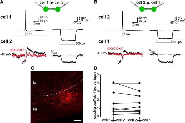
Type 2 GABAergic cells are synaptically and electrically interconnected. A, Paired recordings were made from Type 2 GFP+ cells and synaptic connections between them tested. AP evoked in the presynaptic Type 2 cell (cell 1, top left) by a brief depolarizing current step (middle), and synaptic currents recorded in the postsynaptic neuron (Vh= −45 mV; cell 2, bottom left). APs in the presynaptic cell evoked an outward current in the postsynaptic cell (black trace, bottom left), which could be blocked by picrotoxin (100 μm, red trace). In addition to the uIPSC, a transient inward current was detected in the postsynaptic cell 2 (arrow) that occurred simultaneously with APs in the presynaptic cell. Hyperpolarization of cell 1 (top right) by a negative current step injection (middle right) evoked a hyperpolarization in cell 2 (bottom right). B, Chemical and electrical synapses between Type 2 cells were reciprocal in 43% (3 of 7) and 100% (10 of 10) of connected pairs, respectively. Similar chemical and electrical synaptic connections were also observed from cell 2 to cell 1. APs in cell 2 (top left) evoked uIPSCs in cell 1 (bottom left, black) that were blocked by picrotoxin (bottom left, red). Furthermore, sustained hyperpolarization of cell 2 (top right) evoked a smaller but simultaneous hyperpolarization in cell 1 (bottom right), confirming electrical coupling. C, Electrical synapses occurred between Type 2 cells that were in close proximity of each other (maximum of 50 μm distance between cell bodies, n = 10), but absent between those that were >50 μm apart (n = 2). Photomicrograph represents the biocytin-filled pair of cells presented in A and B. Dashed line indicates the border of the molecular (lm) and cellular layers (lc). D, Strength of electrical coupling was the same in both directions. Graph represents coupling coefficient of electrically connected pairs of Type 2 cells, measured as the ratio of peak hyperpolarization in the postsynaptic cell to that in the presynaptic cell (paired Student's t test, p > 0.05). Scale bar, 50 μm.

