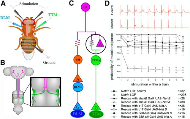
Figure 1.
Physiological recording from the GFS and an analysis of circuit response to repetitive stimulation. A, Schematic of the electrode placement for physiological recording. B, The circuit is composed of a pair of presynaptic giant interneurons, the GF (magenta) and the postsynaptic motor neurons (TTMn, green; DLMn, not shown). The GF projects its axons into the mesothoracic neuromere, where it synapses with the TTMn (inset). C, Wiring diagram of the circuit. The electrical synapses are shown as resistors and the chemical synapses as triangles (“G” for glutamate and “A” for acetylcholine). The giant synapse between the GF and TTMn is composed of both chemical and electrical components. D, The circuit's response at high-frequency stimulation for control, mutant, and rescue experiments. Control specimens (top red trace) responded 1:1 for every stimulation at 100 Hz. Netrin LOF mutants (bottom red trace) did not follow at 100 Hz stimulation. In each animal we tested, we calculated the probability of recording a muscle response on stimulus 1–10 within a single train of stimuli. Netrin heterozygous controls followed nearly 100% for every stimulus within a train. The Netrin LOF mutants exhibited response depression with the probability of response rate falling to ∼45% on stimuli 2–10 of a train. We shifted the following frequencies of the circuit back into near normal ranges in the rescue experiments in which we expressed Netrin under the control of various GAL4 drivers.

