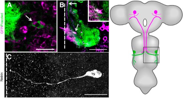
Figure 6.
Netrin and Frazzled expression in GFS. A–C, Netrin and Frazzled production in the GFS between 9% and 27% of PD. A, TTMn cell body (arrow) was labeled with UAS-GFPCD8 (magenta) and Frazzled was labeled with anti-Frazzled antibody (green). Figure is representative of 27% of PD. Scale bar, 20 μm. B, Frazzled signal (green) was also detected and colocalized (white) in the GF axon (magenta) terminal (arrow) of the same specimen in the mesothoracic neuromere. Scale bar, 50 μm. Panels are single images from a z-stack with 0.5 μm steps. C, anti-Netrin antibody was detected on the TTMn medial dendrite and in the cell body. Figure is representative of 18% of PD. The Lateral dendrite of TTMn did not exhibit anti-Netrin signal because it had not projected from the TTMn at this time in PD. Panel is a compressed z-stack. Schematic of adult CNS with box indicates region examined. Scale bar, 20 μm. Dotted lines indicate location of the midline.

