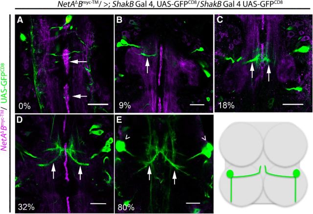
Figure 7.
Development of TTMn's medial dendrite during NetrinB expression in midline glia. During PD, we labeled tethered Myc-tagged NetrinB and expressed UAS-GFPCD8 in TTMn with the shakB-GAL4 driver. A, At 0% of PD, the NetrinB labeling (magenta) was detected on the midline glia (arrows) and longitudinal tracks. B, By 9% of PD, NetrinB signal was observed at the midline and the medial dendrite of TTMn (green) extended (arrow) toward the midline. C, At 18% of PD, the TTMn filopodia (arrows) were at the midline. D, At 32% of PD, the medial dendrite (arrows) was anatomically mature. E, NetrinB production at the midline was no longer observed by 80% of PD. The panels focus on the development of TTMn's medial dendrite. Therefore, the TTMn cell bodies (arrowheads) were absent from the plane of focus in all panels except for E. All panels are single images from a z-stack with 0.5 μm steps. Schematic illustrates TTMns (green) in relation to prothoracic and mesothoracic neuromeres. Scale bar, 20 μm.

