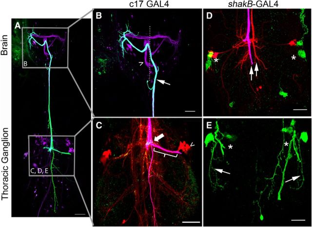
Figure 9.
Expressing tethered NetrinB in glia or the TTMn did not rescue Netrin LOF defects. A–C, When we expressed tethered NetrinB in midline glia with the c17 driver, Netrin LOF mutants were not rescued. B, The GFs displayed midline crossing defects in the brain (arrow and open arrowhead). In this example, both GFs crossed the midline in the brain, but only one GF projected toward the mesothoracic neuromere (arrow). C, The GF that reached the thorax displayed multiple branches (fuchsia) and Innexin (green) was undetectable in the GF axon terminal (bracket). The TTMn cell body (open arrowhead) and dendrites were visible due to dye coupling (red neurobiotin). The TTMn did not synapse with the GF in its normal synaptic area (bracket), but instead at the GF-PSI synaptic region (large arrow). In this region, Innexin staining was observed. Coloring in C is different from A and B because there are three colors used for labeling in C, whereas only two colors are used in A and B. D, E, When we expressed tethered NetrinB in the TTMn with the shakB-GAL4 driver, Netrin LOF mutant defects were not rescued. D, GFs (fuchsia and red) did not make normal terminals or project toward their synaptic targets (arrows). TTMns (green) did not produce dendritic trees (asterisks). E, Some TTMns did project lateral dendrites (arrows), but did not project medial dendrites toward the midline (asterisks). Scale bar, 20 μm.

