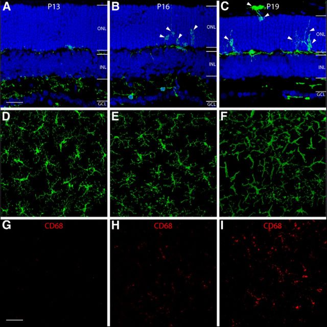
Figure 2.
The initiation of microglia activation and migration in rd10 retinas. In retinal sections (A–C), GFP-expressing microglia are observed to migrate into the inner portion of the ONL at P16 (B, arrowheads), and infiltrate the entire ONL by P19 (C, arrowheads). In retinal whole mounts (D–I), GFP-expressing microglia in the OPL are observed to have a ramified morphology at P13 (D). At P16, microglia begin to change their morphology by retracting their processes (E), and turn into an amoeboid shape with few processes by P19 (F). CD68 staining (G–I), a marker for microglia activation, first appeared at P16 (H) and became more intense by P19 (I). OPL, outer plexiform layer; IPL, inner plexiform layer; INL, inner nuclear layer; GCL, ganglion cell layer. Scale bars: A–C, 20 μm; D–I, 40 μm.

