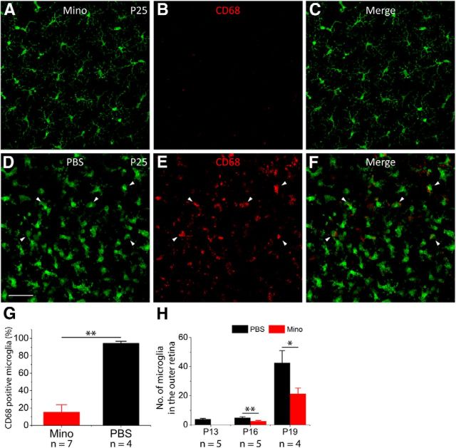
Figure 3.
Minocycline treatment reduces microglia activation and infiltration in rd10 retinas. A–C, At P25, microglia maintain a ramified morphology in minocycline (Mino)-treated rd10 retinas (A), and CD68 staining is dramatically reduced (B). D–F, At P25, microglia show an amoeboid morphology with few processes in PBS-treated rd10 retinas (D), and CD68 staining is intense and widespread (E). G, The percentage of CD68-positive microglia over total microglial cells. H, Quantification of microglial cells that migrated into the outer retina in PBS-treated and minocycline-treated animals at three time points before P25. Results are presented as mean ± SD; *p < 0.05, **p < 0.01, n = number of retinas in each group. Scale bar, 20 μm.

