
Figure 2.
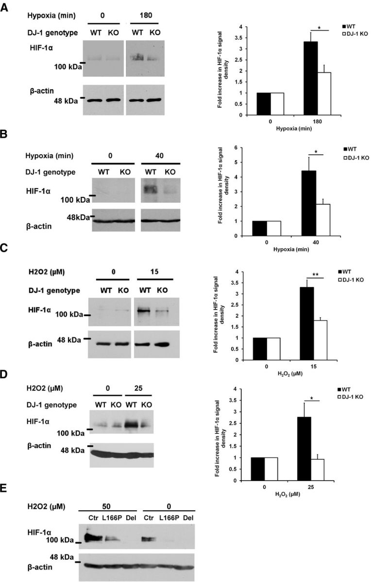
Functional link between DJ-1 and VHL. A, B, WT or DJ-1 KO cortical neurons (A) or MEFs (B) were incubated in normal condition or in hypoxia (1% oxygen) for 180 min (neurons) or 40 min (MEFs). C, Cortical neurons were treated with 15 μm H2O2 for 2 h. D, WT and DJ-1 KO MEFs were also exposed to 0 or 25 μm H2O2 for 24 h. Cells were lysed and HIF-1α protein level was assessed with Western blot using HIF-1α antibody. Densitometry analysis of HIF-1α protein for each experiment is demonstrated in the right side of each panel. Data are the mean ± SEM from three independent experiments. *p < 0.05; **p < 0.01, ANOVA with Tukey's post-test. E, Lymphoblasts extracted from two PD cases, one with a deletion in DJ-1 (Del) and one with a point mutation in DJ-1 (L166P), or from control (Ctr) individual were exposed to 50 μm hydrogen peroxide for 24 h and their HIF-1α response was examined with Western blot analysis using HIF-1α antibody.