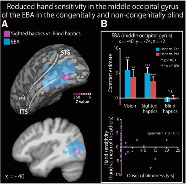
Figure 6.
Reduced hand sensitivity in the middle occipital gyrus of the EBA in the blind subjects. A, Brain regions showing greater hand-sensitive activation in the sighted group than in the blind group (congenital and noncongenital) are shown in purple on a surface-rendered MRI (upper row) and a sagittal section (lower row) of an MRI averaged across the subjects. We used data from both the congenitally and noncongenitally blind subjects in this analysis. The activation was thresholded at p < 0.05, corrected for multiple comparisons over the EBA in each hemisphere, with the height threshold set at Z > 2.58. STS, Superior temporal sulcus; ITS, inferior temporal sulcus. See Table 7 for more information. B, Top, The contrast estimate (relative to each object class) extracted from the peak coordinate in the three conditions. Asterisks indicate the results of one-sample t tests. Bottom, Weak relationship between hand sensitivity and the age at onset of blindness. Data are presented as the mean ± SEM of 28 blind and 28 sighted subjects.

