
Figure 1.
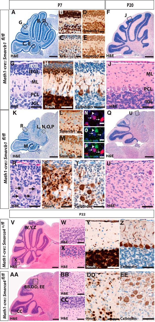
Loss of Smarcb1 in cerebellar granule cells results in a severe reduction of the cerebellar size. Cerebellar histopathology of control mice is normal (A, F) with the typical cortical layering (G, J) as visualized by H&E stains of P7 and P20 sagittal sections. Cre is strongly expressed in rostral parts (B), but less extensive in caudal lobes of control mice (C), and nuclear staining of Smarcb1 is retained in all control cells (D, E). In contrast, Math1-cre::Smarcb1fl/fl mice display a severe phenotype in the Cre-expressing rostral parts of the cerebellum. At P7 and P20, a loss of normal architecture and blurred cortical layers are apparent (K, Q and R, U, respectively), which is caused by a loss of Smarcb1 protein expression in rostral parts and, to a lesser extent, in caudal parts of the cerebellum (L, M). Smarcb1 is particularly lost in Cre-positive cells (N–P), the overall number of NeuN-positive cells is severely decreased in Math1-cre::Smarcb1fl/fl mice, and Purkinje cells are randomly distributed within the cerebellar cortex as compared with controls (H, I, S, T). With respect to histomorphology (V–X and AA–CC) and expression of NeuN and Calbindin (Y, Z, DD, EE), Math1-cre::Smarca4fl/fl mice display changes that are similar, if not identical, to Math1-cre::Smarcb1fl/fl mice. oEGL, Outer external granular layer; iEGL, inner external granular layer; ML, molecular layer; PCL, Purkinje cell layer; IGL, inner granular layer. Scale bars: A, K, 250 μm; B–E, L, M, 50 μm; F, Q, 500 μm; N–P, 10 μm; G–J, R–U, W–Z, BB, CC, DD, EE, 100 μm; V, AA,1 mm.