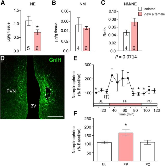
Figure 2.
Effects of a female presence on diencephalic NE and NM concentration, NM/NE ratio, and NE release in the PVN of males. A–C, Effects of a female presence on diencephalic NE concentration (A), NM concentration (B), and NM/NE ratio (C). Data are presented as mean ± SEM. Numbers in the bars denote the number of birds analyzed. Unpaired t test, *p < 0.05 versus the isolated group. D, Immunostaining for GnIH and the placement of the microdialysis probe within the PVN in a frontal section, which is shown in a dotted line. Scale bar, 500 μm. 3V, Third ventricle. E, Extracellular NE in the PVN during BL, FP, and PO. One-way repeated-measures ANOVA followed by Dunnett's multiple comparisons test, †p = 0.0748, *p < 0.05 versus just after seeing a female. n = 6. F, Mean changes in NE released in the PVN during BL, FP, and PO. One-way ANOVA followed by Tukey's multiple comparisons test, *p < 0.05 versus BL and PO.

