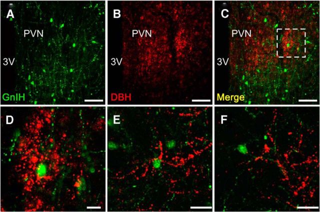
Figure 4.
Noradrenergic neurons project to GnIH neurons. Example of double-immunofluorescent staining for GnIH and DBH immunoreactivities in the PVN from two male quail. Representative fluorescent photomicrographs showing GnIH (A; green) and DBH (B; red). Merged image of A and B (C). D shows higher magnification of the boxed area in C. Another example of GnIH perikarya and dendrites in the PVN (E and F are from the same bird). Similar results were obtained in repeated experiments by using three different birds. 3V, Third ventricle. Scale bars: A–C, 50 μm; D, 10 μm; E, F, 20 μm.

