
Figure 3.
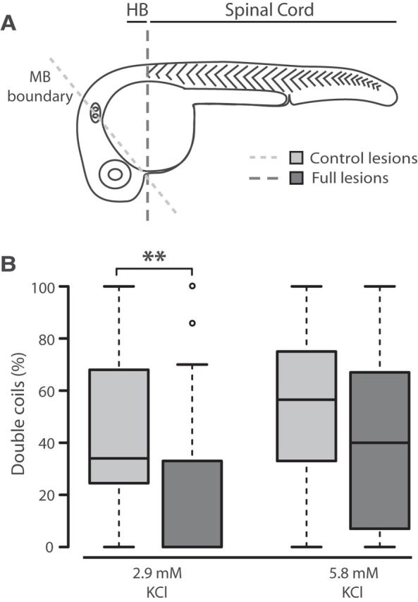
Double coils require a descending excitatory drive from the hindbrain. A, Diagram representing the site of control lesions at the midbrain (MB) boundary to leave the hindbrain (HB) intact, and full lesions at the caudal hindbrain to isolate the spinal cord. B, Box plot showing the proportion of double coils at 26 hpf in control (gray bars) and fully lesioned embryos (dark bars) in normal 2.9 mm KCl (left, Nctrl = 32, Nlesion = 41) and elevated 5.8 mm KCl (right, Nctrl = 22, Nlesion = 25). **p < 0.01.