
Figure 5.
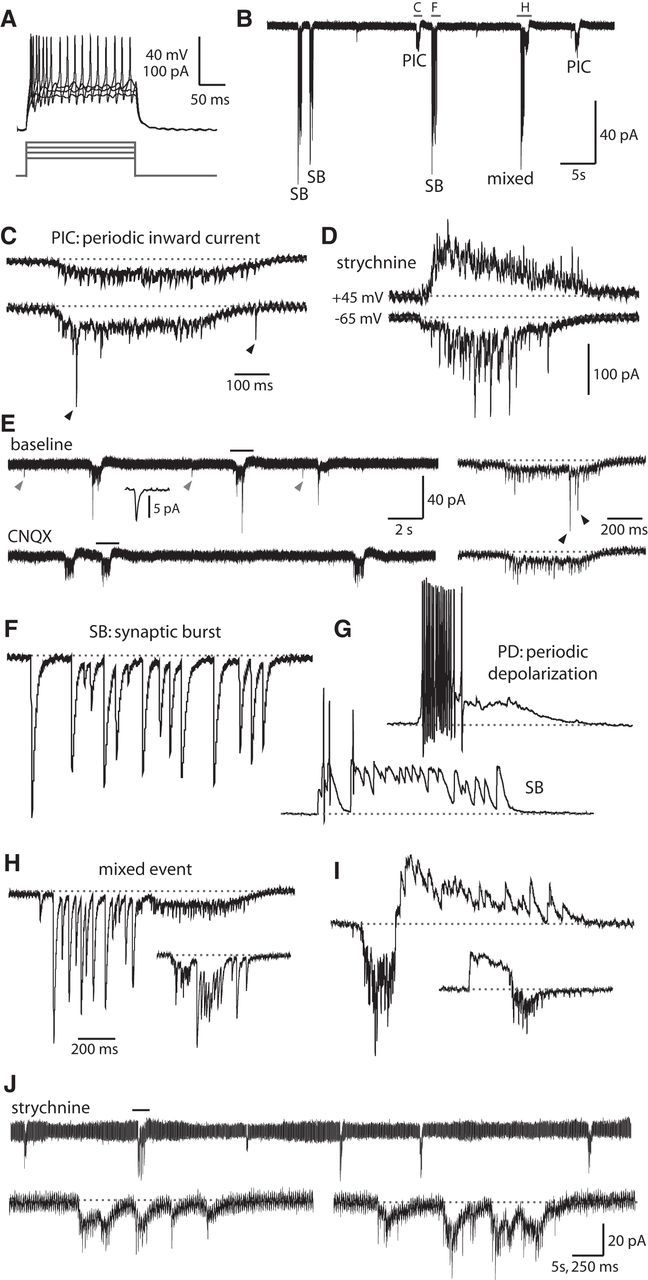
Motoneurons show mixed electrical and synaptic events corresponding to glutamate-dependent fictive double coils. A, Representative current injections showing burst firing of action potentials in embryonic MNs. Top, Current-clamp recording from neurons. Bottom, Current steps. B, Patterned activity in a 25 hpf embryo includes gap junction-driven PICs, SBs, and mixed events. Holding potential in this trace and all others is −65 mV (unless otherwise indicated). Events in the indicated region are expanded in panels C, F, and H. C, PICs are low-amplitude inward currents produced by the electrical coupling of neurons that often coincide with synaptic glutamatergic events. Top, PIC (from region indicated in B) that shows no glutamatergic peaks. Bottom, PIC in the same MN that does show synaptic glutamatergic events (arrowheads). Baseline is shown as a dotted gray line for reference in this and subsequent figures. Vertical scale same as for B, but note different time scale. D, Examples of PICs under voltage clamp seen in an older embryo (27 hpf) with glutamatergic events of increased amplitude and frequency. These recordings were done in the presence of 5 μm strychnine. Some of the currents are reversed at a positive holding potential of 45 mV (top) compared with a normal holding potential (−65 mV, bottom), corresponding to purely glutamatergic events. Time scale same as in C, but note different vertical scale. E, Excerpt from a voltage-clamp recording in a 26 hpf embryo showing three examples of PICs at baseline (left, top) and in the presence of 10 μm CNQX (left, bottom). Small amplitude peaks occurring between events resembling glutamatergic mEPSCs are highlighted with gray arrowheads, and an average of several peaks (n = 19) is shown in the inset at 100 times expanded time scale. Regions indicated with a black bar are shown on a 10 times expanded time scale at the right for each condition. Glutamatergic peaks occurring during a PIC are indicated with black arrowheads. F, Synaptic bursts (from region indicated in B) are strychnine-sensitive glycinergic currents of large amplitude with a similar duration to PICs. G, Representative whole-cell current-clamp recordings from a primary MN in a 26 hpf embryo. Resting membrane potential of the neuron is −60 mV, scale same as in C. Top, Spontaneous periodic depolarization (PD) triggers burst firing of action potentials. Bottom, Glycinergic SB can also trigger action potentials. H, Representative whole-cell voltage-clamp recordings of a mixed events (top trace from region indicated in B) during which a PIC (with or without glutamatergic peaks) and an SB occur in close (subsecond) succession in either order. Vertical scale for uppermost trace same as in B, but note different time scale. Inset, Scaled down by 50%. I, Examples of mixed events from the same MN as in G under voltage clamp at an intermediate holding potential of −20 mV (more positive than the chloride reversal potential of ∼−35 mV) showing that glycinergic currents, but not electrical and glutamatergic currents, are reversed. Scale for top trace and inset same as for H. J, Top, A 100 s excerpt of an MN voltage-clamp recording in a 26 hpf embryo in the presence of 5 μm strychnine showing that glycinergic synaptic bursts are absent, but gap junction-driven PICs remain. Bottom, left, An expanded view of the region indicated in the top trace showing an event with many individual electrical PICs occurring closely together in time and resembling a fictive multiple coil. Bottom, right, Example from a different MN.