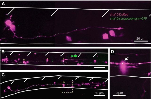
Figure 8.
Embryonic spinal CiD neurons have putative synapses that contact caudal primary MNs and other CiDs. A, Representative example of a DsRed-expressing CiD neuron in a 24 hpf embryo with distinct synaptophysin-GFP puncta along the length of the axon. Spinal cord and somite boundaries are outlined in white. B, A CiD neuron (white asterisk) in a 27 hpf embryo has an axon spanning four somites and synaptophysin-GFP puncta that contact caudal CiDs (yellow asterisks) as well as a primary MN (yellow m). Scale bar, same as for C. C, A rostral CiD neuron in a 28 hpf embryo has an axon spanning 5–6 somites and contacts a primary MN. D, High-resolution single 1 μm confocal plane showing the putative synaptic contact (arrow) between the CiD and MN from the boxed area in C.

