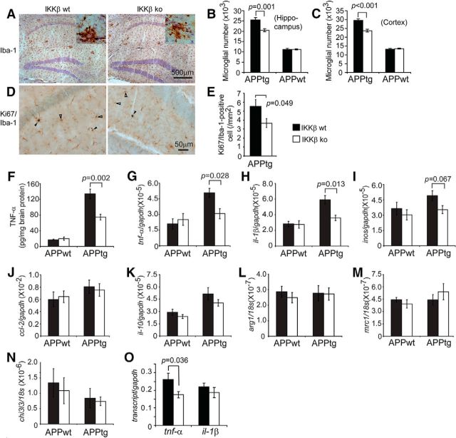
Figure 5.
Deficiency of IKKβ in myeloid cells reduces neuroinflammatory activation in APP-transgenic mice. Six-month-old APP-transgenic mice (APPtg) and their non-APP-transgenic littermates (APPwt) were tested for inflammatory activation. Microglial cell numbers were estimated with stereological methods after immunohistochemical staining of Iba-1 (A; in brown). Proliferating microglia were identified by double staining of Iba-1 and Ki67, which appear in blue nucleus and brown cytoplasm (D; marked with closed arrowheads; pure Iba-1+ cells are marked with open arrowheads). The numbers of Iba-1+ cells were significantly smaller in APPtg mice with ablation of myeloid IKKβ (IKKβko) than in those with normal IKKβ expression (IKKβwt). However, IKKβ ablation did not affect the number of Iba-1+ cells in APPwt mice (B, C; one-way ANOVA; n = 6 per group). The number of Iba-1/Ki67 double-positive cells was significantly smaller in APPtg/IKKβko than in APPtg/IKKwt mice (E; t test; n = 6 per group). TNF-α protein concentration in brain homogenates derived from APPtg and APPwt mice was determined by ELISA (F; one-way ANOVA; n ≥ 6 per group). Inflammatory gene transcripts in the brain (G–N) and in isolated microglia from 6-month-old APPtg mice (O) were measured by quantitative RT-PCR. Both TNF-α protein expression and the number of tnf-α and il-1β transcripts in APPtg brains, but not in the APPwt control brains, were significantly reduced by IKKβ ablation in myeloid cells (F-H; one-way ANOVA; n ≥ 9 per group). Accordingly, the number of tnf-α transcripts in APPtg microglia was also significantly reduced by IKKβ ablation (O; t test; n = 3 per group).

