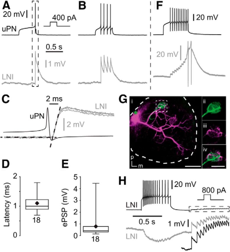
Figure 1.

Type I LNs receive fast, excitatory input from uPNs. A, B, Single presynaptic action potentials in the uPN induced fast, transient short-latency EPSPs in the postsynaptic type I LN. C, Higher magnification of the frame in A showing fast synaptic input, with two additional fast EPSPs from the same neuron. The black dotted lines represent linear fits of the resting membrane potential and the rising phase of an EPSP. Their point of intersection was defined as the onset of the EPSP. D, Latency between the uPN spikes and EPSP in type I LNs. E, EPSP amplitudes in type I LNs that were elicited by single uPN action potentials. F, During high-frequency trains of action potentials the fast EPSPs can reach the action potential threshold (action potentials clipped). Gi, Morphology of the recorded uPN (green) and type I LN (magenta) revealed by staining of each neuron via the recording pipette. The green asterisk marks the position of the uPN soma that was lost during processing. Scale bar, 100 μm. ii–iv, Higher magnification of frame i showing neurites of both neurons in the same glomerulus. m, Medial; p, posterior. Scale bar, 50 μm. H, Recording from two type I LNs showing coincident EPSPs presumably due to dyadic input from a uPN. The black trace in the dotted box is enlarged underneath.
