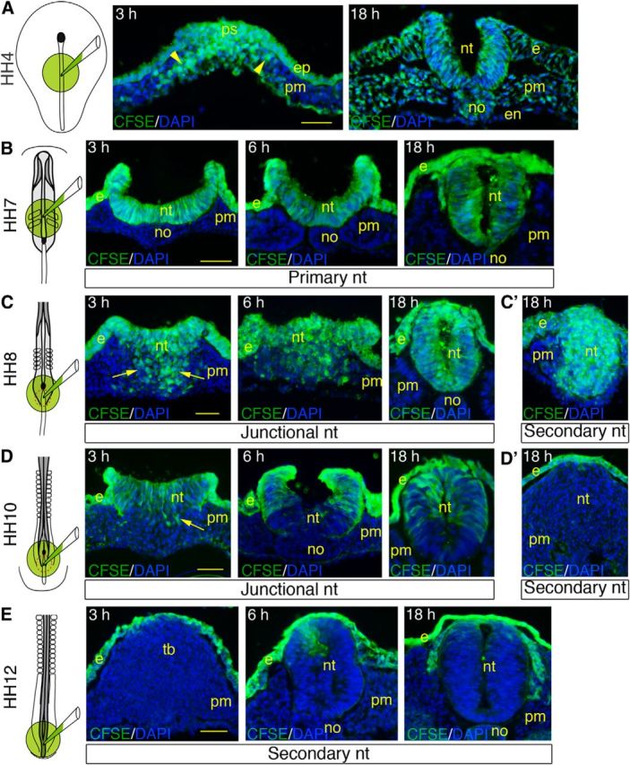
Figure 3.

CFSE tracing of neural progenitors in the NSB. CFSE and DAPI visualization on cross-sections through chick embryos labeled with CFSE at the level of the primitive streak at HH4 (A), the primary neural tube at HH7 (B), the NSB at HH8 (C) and HH10 (D), and the secondary neural tube at HH12 (E). Embryos were labeled for1 h, rinsed, and further incubated during 3, 6, and 18 h before analysis. Far left panels, Embryonic area where the dye was applied. A, Sections through the primitive streak (left) and the anterior trunk (right). B, Sections though the primary neural tube. C, D, Sections through the mid-portion of the NSB (left and middle) and the junctional neural tube (right). C′, D′, E, Sections through the secondary neural tube. Arrowheads point at fluorescent cells ingressing in the primitive streak and migrating laterally; arrows point to fluorescent cells dispersed in the space underneath the superficial layer at the level of the NSB. e, Ectoderm; en, endoderm; ep, epiblast; no, notochord; nt, neural tissue; pm; paraxial mesoderm; ps, primitive streak; tb, tail bud. Scale bar, 50 μm.
