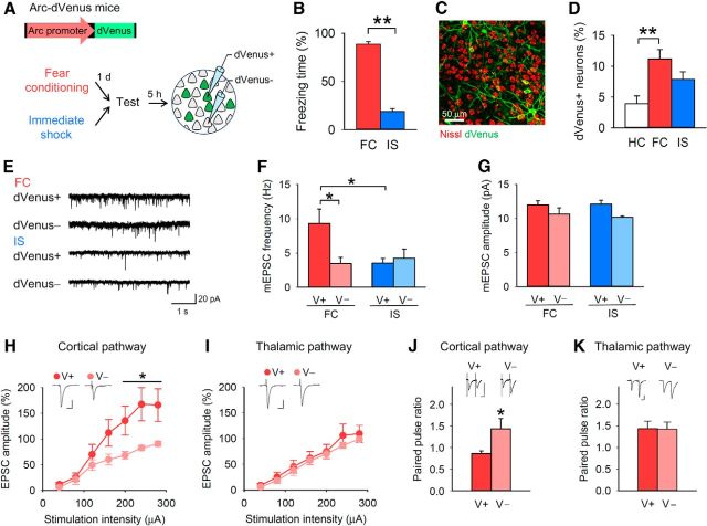
Figure 1.
Contextual fear conditioning induces presynaptic potentiation in BLA neurons recruited into a fear memory trace. A, Experimental procedure. B, The FC group showed longer freezing time during the test than the IS group. C, A representative fluorescence image of BLA neurons with dVenus. D, Re-exposure to the conditioning context increased the proportion of dVenus+ neurons in the BLA (one-way ANOVA, F(2,18) = 6.7, p = 0.0068; Tukey's test, home cage (HC) vs FC, p = 0.0050). E, F, dVenus+ (V+) neurons of the FC group had a higher mEPSC frequency than dVenus− (V−) neurons of the FC group and dVenus+ neurons of the IS group (n = 10 dVenus+ and 10 dVenus− neurons per behavioral group from 14 mice in the FC group and 12 mice in the IS group; two-way ANOVA, F(1,36) = 4.9, p = 0.033; dVenus+ (FC) vs dVenus− (FC), p = 0.042; dVenus+ (FC) vs dVenus+ (IS), p = 0.024). G, There was no significant group × dVenus interaction of mEPSC amplitude (F(1,36) = 0.33, p = 0.57). H, I, The EPSC amplitude of the dVenus+ neurons in the FC group was higher than that of the dVenus− neurons in the FC group in the cortical but not thalamic pathway (n = 7–10 neurons from 5 mice; cortical pathway, repeated-measures ANOVA, F(6,108) = 4.5, p = 4.1 × 10⁻⁴). Calibration: 20 ms, 100 pA. J, K, The PPR in dVenus+ neurons in the FC group was lower than that of dVenus− neurons in the FC group in the cortical but not thalamic pathway (n = 7–11 neurons from 7 mice; cortical pathway, Student's t test, t(11) = 2.3, p = 0.041). Calibration: 20 ms, 50 pA. **p < 0.01, *p < 0.05.

